Blog

Your blog category

penile doppler test in hyderabad

Is Poor Blood Flow Causing Erection Problems? Understanding Penile Doppler Test in Hyderabad

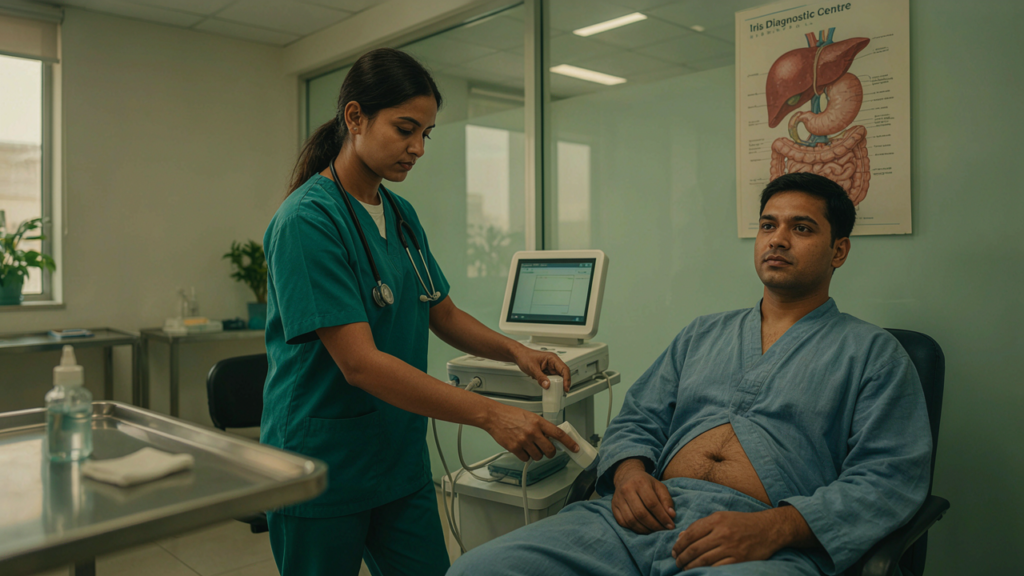
Erection problems can affect a man’s confidence, relationships, and overall quality of life. Although stress and anxiety can contribute to erectile dysfunction, poor blood circulation is another important cause. When the blood vessels cannot deliver enough blood to the penis or blood does not remain trapped inside it, achieving or maintaining an erection may become difficult. Understanding the cause is the first step towards appropriate care. A Penile Doppler Test in Hyderabad is a specialised ultrasound examination that helps doctors assess blood flow in the penile blood vessels. It can provide useful information about possible circulation-related causes of erectile dysfunction and help doctors decide what further evaluation or treatment may be needed. At Iris Diagnostics, patients can seek diagnostic guidance to understand their testing requirements and make informed decisions about their health. If you have been experiencing persistent erection difficulties, learning about a Penile Doppler Test in Hyderabad may help you understand how this examination works and when a doctor might recommend it. What Is Erectile Dysfunction? Erectile dysfunction (ED) is the repeated difficulty in achieving or maintaining an erection firm enough for sexual activity. Occasional erection difficulties can happen because of tiredness, stress, anxiety, or temporary health changes. However, when the problem continues or becomes frequent, it is important to identify the underlying cause. A Penile Doppler Test in Hyderabad helps assess blood flow, which is essential for a healthy erection. An erection depends on several processes working together: the brain sends signals, nerves carry messages, blood vessels deliver blood, and penile tissues help maintain firmness. Hormones and emotional well-being also play important roles in erectile health. If any of these processes is affected, erection difficulties may occur. Common causes of erectile dysfunction include: A doctor may recommend a Penile Doppler Test in Hyderabad when the symptoms suggest a possible blood vessel problem or when more information about penile circulation is needed. Can Poor Blood Flow Cause Erection Problems? Yes. Healthy blood circulation is essential for achieving and maintaining an erection. During sexual stimulation, the blood vessels supplying the penis relax and widen, allowing more blood to enter the erectile tissues. These tissues expand and help restrict blood from flowing out too quickly. When this process does not work properly, an erection may be weak, short-lasting, or difficult to achieve. There are two main blood-flow problems doctors may investigate. 1. Reduced Blood Flow Into the Penis The arteries carry oxygen-rich blood to the penile tissues. If these blood vessels become narrowed or do not widen properly, the penis may not receive enough blood during sexual stimulation. Conditions such as diabetes, high cholesterol, high blood pressure, and atherosclerosis can affect blood vessels. Smoking can also damage the lining of the arteries and increase vascular risks. A Penile Doppler Test in Hyderabad can help assess arterial blood flow and identify findings that may suggest reduced blood supply. 2. Difficulty Keeping Blood Inside the Penis An erection requires more than blood entering the penis. The blood must also remain within the erectile tissues long enough to maintain firmness. If the mechanisms that restrict blood outflow do not function effectively, a man may achieve an erection but lose firmness too quickly. This may be described as a possible veno-occlusive dysfunction. A penile Doppler examination can provide information about blood-flow patterns that help doctors investigate this possibility. However, results must be interpreted alongside symptoms, examination findings, and other clinical information. What Is a Penile Doppler Test? A Penile Doppler Test in Hyderabad is a specialised ultrasound examination that evaluates blood circulation in the penis. It combines conventional ultrasound imaging with Doppler technology, which measures the movement and direction of blood through blood vessels. Unlike an ordinary external examination, this test allows the doctor to assess blood-flow patterns within the penile arteries and veins. The examination may help doctors investigate: The test does not diagnose every possible cause of erectile dysfunction. For example, hormonal problems, anxiety, nerve disorders, and medication-related effects may require different investigations. A Penile Doppler Test in Hyderabad is therefore one part of a broader medical assessment rather than a complete assessment of every aspect of sexual health. How Does a Penile Doppler Test Work? Understanding the procedure can help reduce uncertainty before your appointment. The examination is generally performed using ultrasound equipment and a handheld probe. Depending on the clinical question, the doctor may recommend a basic examination or a dynamic study that evaluates blood flow following medication administered under medical supervision. Step 1: Medical Assessment Before the scan, the doctor may ask about your symptoms, medical history, medications, diabetes, blood pressure, smoking habits, and previous treatments for erectile dysfunction. This information helps determine whether the examination is appropriate and how the findings should be interpreted. Step 2: Ultrasound Examination You will generally lie comfortably on an examination table. A suitable ultrasound gel is applied to the area, and the sonographer or doctor moves the probe over the penis to obtain images of the relevant blood vessels. The ultrasound helps visualise penile structures and assess blood movement. Step 3: Blood-Flow Measurements Doppler technology measures blood-flow characteristics, including the speed and direction of circulation. Measurements can help the doctor evaluate whether arterial inflow appears adequate and whether blood is being retained appropriately. Step 4: Medication-Assisted Assessment, If Required For some patients, a doctor may recommend an injection of a medicine into the penis to produce an erection during the examination. This allows blood flow to be evaluated under stimulated conditions. Not every patient requires this step. The decision depends on the clinical indication and the protocol followed by the treating specialist. If medication is used, the procedure must be supervised by trained medical professionals. The doctor should explain the benefits, possible discomfort, side effects, and precautions before proceeding. Step 5: Review of Findings After the examination, the images and measurements are reviewed. The findings are interpreted alongside the patient’s symptoms and medical history. If you are considering a Penile Doppler Test in Hyderabad, ask the diagnostic centre or referring doctor whether …


Is Poor Blood Flow Causing Erection Problems? Understanding Penile Doppler Test in Hyderabad Read More »

penile doppler test in hyderabad

Erection Problems Despite Treatment? When Is a Penile Doppler Test Recommended?

Erection problems despite treatment can be frustrating and may affect a man’s confidence, relationships, and overall quality of life. Although erectile dysfunction (ED) is common, persistent symptoms should not be ignored, particularly when prescribed medication does not produce the expected improvement. Identifying the underlying cause is an important step toward choosing an appropriate treatment plan. A penile Doppler ultrasound is a specialized imaging investigation that evaluates blood flow within the penis. Doctors may recommend it when erectile dysfunction does not respond adequately to conventional treatment or when a vascular problem is suspected. If you are searching for a penile doppler test in hyderabad, understanding the procedure, its benefits, and when it may be needed can help you make informed decisions with your healthcare provider. At Iris Diagnostic, patients can explore available diagnostic services and discuss appropriate investigations with their referring doctor. The decision to undergo a penile Doppler examination should always be based on clinical assessment rather than symptoms alone. What Is Erectile Dysfunction? Erectile dysfunction is the persistent difficulty in achieving or maintaining an erection firm enough for satisfactory sexual activity. It can occur occasionally because of stress, tiredness, or temporary health concerns. However, when the problem becomes frequent or continues over time, medical evaluation may be necessary. An erection depends on several interconnected processes involving the brain, nerves, hormones, blood vessels, and penile tissues. Sexual stimulation triggers signals that relax smooth muscles and increase blood flow into the erectile tissues. At the same time, the body’s natural mechanisms help retain blood within the penis to maintain rigidity. If any part of this process is affected, erectile difficulties may develop. Common contributing factors include: Because erectile dysfunction can have several causes, treatment that works for one person may not work for another. This is why identifying the underlying problem is often more useful than repeatedly changing medication without medical supervision. Why Do Erection Problems Continue Despite Treatment? Many men initially receive oral medication or lifestyle recommendations to manage erectile dysfunction. These approaches can be effective, but not everyone experiences satisfactory results. If erections remain weak or difficult to maintain despite treatment, a doctor may reassess the diagnosis, medication use, associated health conditions, and possible vascular causes. 1. Reduced Blood Flow to the Penis An erection requires sufficient blood to enter the penile arteries. When these arteries become narrowed or do not deliver adequate blood, achieving a firm erection may become difficult. Conditions such as diabetes, hypertension, high cholesterol, and atherosclerosis can affect blood vessel health. In selected cases, a penile doppler test in hyderabad may help doctors investigate whether reduced arterial blood flow contributes to persistent erectile dysfunction. 2. Difficulty Retaining Blood During an Erection An erection also depends on the ability of penile tissues to restrict blood from flowing out too quickly. When the normal blood-retention mechanism does not function effectively, a man may achieve an erection initially but struggle to maintain it. This is sometimes described as veno-occlusive dysfunction. A penile Doppler examination can help assess blood-flow patterns that may suggest an issue with this mechanism. However, the findings must be interpreted alongside the patient’s symptoms and the technical details of the examination. 3. Diabetes and Metabolic Conditions Diabetes can affect both blood vessels and nerves. Over time, these changes may interfere with the body’s ability to produce and maintain an erection. Men with diabetes may require a comprehensive evaluation that includes glucose control, blood pressure, cholesterol, medication review, and other relevant investigations. A doctor may consider a penile doppler test in hyderabad when vascular assessment is clinically indicated. 4. Hormonal or Neurological Factors Low testosterone and certain neurological conditions can contribute to sexual difficulties. These causes are not necessarily identified through Doppler imaging alone. Depending on the symptoms, a doctor may recommend hormone testing, neurological assessment, or other investigations before deciding whether vascular imaging is appropriate. 5. Psychological and Emotional Factors Stress, performance anxiety, depression, and relationship concerns can affect erections even when penile blood flow is adequate. A normal Doppler result does not mean the symptoms are imaginary. It may simply indicate that a significant vascular problem was not demonstrated during the examination. Psychological, hormonal, neurological, and other factors may still require attention. What Is a Penile Doppler Test? A penile Doppler test, also called penile duplex Doppler ultrasound, is an imaging procedure used to assess blood circulation within the penis. It combines conventional ultrasound imaging with Doppler technology, which evaluates the movement of blood through blood vessels. The examination can provide information about arterial blood entering the erectile tissues and blood-flow patterns associated with maintaining an erection. Doctors may recommend this investigation when they need more information about the possible vascular causes of erectile dysfunction. If you are researching a penile doppler test in hyderabad, it is important to understand that this examination is not routinely required for every man experiencing erection problems. Many patients can be evaluated through medical history, physical examination, medication review, and appropriate laboratory tests. A Doppler examination is generally considered when the results could help clarify the diagnosis or guide further management. What Can a Penile Doppler Ultrasound Detect? Depending on the examination and clinical circumstances, it may help assess: The test does not independently diagnose every possible cause of erectile dysfunction. Its value lies in answering specific clinical questions about penile anatomy and circulation. When Is a Penile Doppler Test Recommended? A doctor may recommend a penile doppler test in hyderabad when persistent symptoms require a more detailed assessment of penile blood flow. The following situations may justify further evaluation. 1. When Oral ED Medication Does Not Work Medication response depends on the underlying cause, correct use, dose, timing, sexual stimulation, and other individual factors. Before recommending additional investigations, a doctor may review whether the medication was used as prescribed and whether any medical conditions or interacting medicines could affect its effectiveness. If erectile difficulties continue despite an appropriate treatment trial, penile Doppler imaging may help investigate suspected vascular problems. 2. When an Arterial Blood-Flow Problem Is …

Erection Problems Despite Treatment? When Is a Penile Doppler Test Recommended? Read More »

Scrotum Scan For infertility

Male Infertility Explained: What Your Scrotal Ultrasound Can Reveal

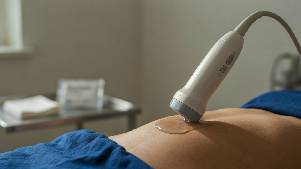
Male infertility is a common reproductive health concern that can affect couples who are trying to conceive. While infertility can have many causes, problems involving the testicles, epididymis, sperm-producing tissues, and blood vessels around the testicles can sometimes contribute to reduced fertility. Identifying these conditions early can help doctors determine the most appropriate treatment or further investigations. One of the useful imaging investigations for evaluating male reproductive health is a Scrotum scan for infertility. Also known as a scrotal ultrasound, this examination uses sound waves to create detailed images of the testicles and surrounding structures. It can help identify abnormalities that may not be obvious during a physical examination. At Iris Diagnostics, advanced diagnostic imaging can support doctors in evaluating scrotal and reproductive health concerns. A Scrotum scan for infertility can provide valuable information when infertility is suspected, particularly when symptoms such as testicular pain, swelling, a lump, or suspected varicocele are present. What Is Male Infertility? Male infertility refers to difficulty achieving pregnancy because of factors affecting the male reproductive system. It may be related to low sperm production, poor sperm movement, abnormal sperm structure, hormonal problems, blockages, testicular conditions, or other reproductive health issues. In some cases, a man may have no obvious symptoms. A person may feel completely healthy while still having an underlying condition that affects sperm production or sperm quality. This is why fertility evaluation often involves a combination of medical history, physical examination, semen analysis, blood tests, and imaging when clinically indicated. A Scrotum scan for infertility can complement these investigations by allowing doctors to examine the physical structures involved in sperm production and transport. What Is a Scrotal Ultrasound? A scrotal ultrasound is a non-invasive imaging examination of the scrotum. It uses high-frequency sound waves to generate real-time images of the testicles, epididymis, surrounding tissues, and blood vessels. Unlike X-rays, ultrasound does not use ionizing radiation. The examination is generally painless and does not require an incision or invasive procedure. A Scrotum scan for infertility may be recommended when a doctor wants to investigate structural or vascular causes that could potentially affect reproductive function. During the examination, an ultrasound transducer is gently moved over the scrotal area using ultrasound gel. The radiologist or sonologist evaluates the appearance, size, position, and internal structure of the testicles and surrounding tissues. Why Is a Scrotum Scan for Infertility Recommended? A Scrotum scan for infertility may be recommended as part of a fertility assessment when there is a possibility of a structural abnormality affecting reproductive function. Doctors may suggest the examination when a patient has: It is important to understand that a Scrotum scan for infertility does not measure sperm count directly. Instead, it evaluates anatomical and vascular structures that may provide clues about conditions associated with male infertility. What Can a Scrotum Scan for Infertility Reveal? One of the major advantages of a Scrotum scan for infertility is its ability to visualize structures that cannot be assessed adequately through external examination alone. The scan may help identify: 1. Varicocele A varicocele refers to enlarged veins within the scrotum, particularly around the testicle. It is one of the conditions commonly considered during the evaluation of male infertility. A Scrotum scan for infertility can help visualize enlarged veins and, when Doppler ultrasound is used, assess blood flow within those vessels. Varicoceles may be associated with changes in the temperature and blood circulation around the testicles. In some individuals, they may be associated with impaired sperm production or quality. Not every varicocele causes infertility, so findings from the scan need to be interpreted alongside symptoms, physical examination, and semen analysis. 2. Testicular Size and Structure The testicles are responsible for producing sperm and testosterone. Changes in testicular size or internal structure may provide useful information during fertility evaluation. A Scrotum scan for infertility allows doctors to assess the testicular tissue and compare the appearance of both testicles. Differences in size, abnormal tissue patterns, or other structural changes may require additional clinical evaluation. 3. Epididymal Abnormalities The epididymis is a coiled structure located behind each testicle where sperm mature and are transported. Inflammation, cysts, or other abnormalities involving the epididymis may sometimes be visible during a Scrotum scan for infertility. These findings can be particularly relevant when fertility problems occur alongside scrotal pain, swelling, or a history of infection. 4. Testicular Cysts Ultrasound can identify fluid-filled structures such as cysts within or around the scrotum. Some cystic findings are harmless and may not affect fertility, while others may require clinical monitoring depending on their location, size, and appearance. A Scrotum scan for infertility helps provide anatomical information that allows the treating doctor to decide whether additional evaluation is necessary. 5. Hydrocele A hydrocele occurs when fluid collects around a testicle. It can cause swelling or a feeling of heaviness in the scrotum. Although a hydrocele does not automatically mean infertility, identifying it during a Scrotum scan for infertility can help explain certain scrotal symptoms and distinguish it from other causes of swelling. 6. Testicular Masses Ultrasound can help identify unusual areas or masses within the testicle. Not every testicular mass is cancerous, but an abnormal finding should always be evaluated by an appropriate medical professional. A Scrotum scan for infertility can therefore be useful when a lump or abnormality is detected during examination. 7. Testicular Torsion and Blood-Flow Changes Doppler ultrasound can evaluate blood flow to the testicles. This can be particularly important when there is sudden and severe testicular pain. Although testicular torsion is an emergency rather than a routine infertility condition, Doppler imaging can help assess blood supply when clinically indicated. A Scrotum scan for infertility involving Doppler assessment may also provide information about blood circulation in cases where vascular abnormalities are suspected. How Does Doppler Ultrasound Help? Doppler ultrasound is an important component of many scrotal ultrasound examinations. It evaluates movement of blood through vessels and can help detect vascular abnormalities. In the evaluation of suspected varicocele, Doppler imaging can help demonstrate dilated veins and changes …

Male Infertility Explained: What Your Scrotal Ultrasound Can Reveal Read More »

FibroScan in Hyderabad

Can FibroScan in Hyderabad Help Detect Liver Damage Early?

can FibroScan in Hyderabad Help Detect Liver Damage Early? Liver health is an important part of overall wellness, but liver damage can sometimes develop silently without clear symptoms. Early evaluation can help people understand their liver condition and take timely steps toward better health. FibroScan in Hyderabad is a modern, non-invasive liver assessment method that can provide useful information about liver stiffness and liver fat. When recommended by a healthcare professional, it can support the early assessment and monitoring of several liver-related conditions. Understanding liver health at an early stage can encourage positive lifestyle changes and appropriate medical follow-up. FibroScan in Hyderabad can be particularly helpful for people with risk factors such as fatty liver, diabetes, obesity, viral hepatitis, metabolic concerns, or abnormal liver test results. What Is FibroScan in Hyderabad? FibroScan in Hyderabad refers to non-invasive liver assessment using FibroScan technology to measure liver stiffness and, where applicable, estimate liver fat through a Controlled Attenuation Parameter (CAP) measurement. The examination uses specialized technology to assess how waves move through liver tissue. The resulting measurement can provide information that may help healthcare professionals evaluate the possibility of liver fibrosis. Unlike an invasive biopsy, FibroScan does not require tissue to be removed from the liver. This makes it a convenient assessment option for suitable patients. Why Early Liver Damage Detection Matters The liver has an impressive ability to perform essential functions and may continue working even when damage is developing. As a result, some liver conditions may not cause noticeable symptoms during their early stages. Early assessment can provide an opportunity to identify potential concerns before they become more advanced. FibroScan in Hyderabad can contribute to this process by providing measurable information about liver stiffness. Early awareness can also motivate people to improve nutrition, physical activity, weight management, and other lifestyle factors that support better liver health. How FibroScan in Hyderabad Supports Early Assessment One of the important advantages of FibroScan in Hyderabad is that it provides a quantitative liver stiffness measurement. Liver stiffness can be associated with fibrosis, which develops when repeated or long-term liver injury results in scar tissue formation. FibroScan can help healthcare professionals assess stiffness without requiring a surgical procedure. However, liver stiffness results should not be considered a diagnosis on their own. Healthcare professionals interpret the measurement alongside medical history, blood tests, symptoms, imaging, and other relevant information. Understanding Liver Stiffness Liver stiffness is commonly reported in kilopascals (kPa). Changes in stiffness may provide useful information about the condition of liver tissue. A higher measurement may be associated with a greater likelihood of fibrosis, but stiffness can also be influenced by factors other than fibrosis. Therefore, professional interpretation is important. FibroScan in Hyderabad can provide a valuable measurement that becomes more meaningful when considered as part of a complete liver assessment. Can FibroScan Detect Liver Damage Early? FibroScan can help assess liver stiffness, which may provide information related to fibrosis. It is particularly useful as a non-invasive tool for evaluating liver health in appropriate patients. It is important to understand that FibroScan does not identify every form of liver damage by itself. Instead, FibroScan in Hyderabad can complement other investigations and help healthcare professionals build a clearer picture of liver health. Early detection depends on the underlying condition, the individual’s risk factors, and the combination of tests used during evaluation. FibroScan in Hyderabad and Fatty Liver Fatty liver is a common liver health concern that can occur when excess fat accumulates in liver cells. It is often associated with metabolic factors such as obesity, diabetes, insulin resistance, and abnormal cholesterol levels. Many people with fatty liver may not experience obvious symptoms. This makes appropriate evaluation and monitoring especially important. FibroScan in Hyderabad can provide CAP measurements that help estimate liver fat and liver stiffness measurements that can support fibrosis assessment. Together, these measurements may offer useful information about different aspects of liver health. The Connection Between Fatty Liver and Fibrosis Fat accumulation does not necessarily mean that a person has advanced liver disease. However, in some individuals, ongoing liver injury can contribute to inflammation and fibrosis. Monitoring can help healthcare professionals understand the overall situation and determine whether additional assessment is needed. For suitable patients, FibroScan in Hyderabad can provide non-invasive measurements that complement blood tests and other diagnostic methods. Role of FibroScan in Liver Fibrosis Assessment Fibrosis refers to the development of scar tissue within the liver. It can occur when the liver experiences repeated or long-term injury. Fibrosis can develop gradually, and symptoms may not always be obvious. Measuring liver stiffness can therefore provide useful information during medical evaluation. FibroScan in Hyderabad may be recommended when a healthcare professional wants additional non-invasive information about the possibility or extent of liver fibrosis. Who May Need Liver Health Assessment? Not everyone needs FibroScan testing. A healthcare professional can determine whether the test is appropriate based on personal risk factors and medical history. People with fatty liver, chronic hepatitis, abnormal liver enzymes, metabolic conditions, or other known liver concerns may be considered for further liver assessment. Individuals with multiple risk factors may also benefit from discussing liver monitoring with their healthcare provider. FibroScan in Hyderabad may be one of the tools considered as part of that assessment. Important Risk Factors for Liver Damage Several factors can increase the risk of liver problems. These may include: Risk Factor Possible Liver Concern Fatty liver Accumulation of fat in liver tissue Obesity Increased metabolic and liver-related risk Diabetes Higher risk of metabolic liver disease Viral hepatitis Long-term liver inflammation Excessive alcohol use Alcohol-related liver injury High cholesterol Associated metabolic risk Certain medications May affect liver health in some individuals Family or medical history May influence individual risk Understanding these factors can encourage proactive and positive health decisions. How FibroScan in Hyderabad Can Support Monitoring Liver assessment is not always about identifying a problem for the first time. It can also involve tracking changes over time. When follow-up testing is clinically appropriate, measurements may be compared with previous results. This …

Can FibroScan in Hyderabad Help Detect Liver Damage Early? Read More »

Scrotum Scan for Infertility

Scrotum Scan for Infertility: Detecting Varicocele and Other Conditions

Male fertility is influenced by several important factors, including healthy sperm production, normal testicular function, proper hormone balance, and the effective movement of sperm through the reproductive system. When a couple experiences difficulty conceiving, a careful and complete evaluation can help identify possible contributing factors. A Scrotum Scan for Infertility is an ultrasound-based examination that can provide valuable information about the testicles, epididymis, scrotal tissues, and blood vessels. It may be recommended when a healthcare professional needs to investigate a possible structural or vascular problem that could be relevant to male reproductive health. The scan uses sound waves to create images of the scrotal structures and does not use ionizing radiation. It may also include Doppler ultrasound, which can provide information about blood flow. This makes it a useful imaging method when conditions such as varicocele or other scrotal abnormalities need to be assessed. What Is a Scrotum Scan for Infertility? A Scrotum Scan for Infertility is a diagnostic ultrasound examination designed to evaluate the structures within the scrotum. The testicles, epididymis, surrounding tissues, and blood vessels can be examined through ultrasound imaging. The examination can provide clear and useful visual information that may not be obtained through a physical examination alone. Ultrasound produces images by sending high-frequency sound waves into the body and recording the echoes that return. Because it does not use X-rays, it is considered a non-invasive imaging technique. When Doppler technology is used, the examination can also show blood movement through vessels. This can be particularly helpful when a doctor suspects abnormal venous circulation or wants to assess a possible varicocele. It is important to understand that a scrotal ultrasound is not a complete infertility test. Instead, it is one useful tool that may support a broader fertility evaluation. Why Is Scrotum Scan for Infertility Important? Male infertility may have several possible causes, and some of them can be associated with conditions involving the testicles or scrotal structures. A Scrotum Scan for Infertility can help healthcare professionals look for specific structural or vascular findings that may require further evaluation. Depending on the clinical situation, ultrasound may help assess: The value of the examination comes from combining its findings with the patient’s medical history, symptoms, physical examination, and other fertility tests. A clear imaging result can provide important information and may help make the overall evaluation more focused and effective. How Male Fertility Can Be Affected Healthy sperm production depends on properly functioning testicular tissue. Hormones, blood supply, temperature regulation, and the reproductive ducts all have important roles. When one or more of these systems is affected, sperm production or sperm function may be reduced. For some men, the problem is related to sperm count, movement, or shape. For others, a structural condition, hormonal issue, infection, or reproductive tract problem may contribute to fertility difficulties. Varicocele is one example of a condition that can be associated with male infertility. It involves enlarged veins that drain the testicle and may be linked with lower sperm quantity or quality in some men. Because male fertility has many possible causes, imaging may be recommended when a structural or vascular concern is suspected. Scrotum Scan for Infertility and Varicocele Detection One of the most important conditions evaluated through scrotal ultrasound is varicocele. A varicocele is an enlargement of veins in the scrotum. It can sometimes cause pain or a feeling of heaviness, but some men may have no noticeable symptoms. During a physical examination, a healthcare professional may be able to identify a varicocele by inspecting and feeling the scrotum. Ultrasound can provide additional information and may help confirm or characterize the condition. Doppler imaging is particularly valuable because it can assess blood flow within the scrotal veins. This information may help the doctor understand whether abnormal circulation is present and whether further evaluation is appropriate. The presence of a varicocele does not automatically mean that it is the sole reason for infertility. Doctors generally interpret the imaging results together with semen analysis, fertility history, physical examination, and other relevant findings. How Does Varicocele Affect Fertility? The exact way varicocele affects fertility is not completely understood. Research and clinical evidence suggest that abnormal venous circulation may affect the testicular environment and may be associated with reduced sperm production or quality in some men. Possible effects may involve temperature regulation, blood flow, and the local environment in which sperm are produced. Not every man with varicocele experiences infertility. Some men may have normal fertility despite having the condition, while others may show abnormalities in semen parameters. This is why identifying a varicocele is only one part of a proper evaluation. Other Conditions a Scrotum Scan for Infertility Can Help Assess Although varicocele is a major reason for scrotal ultrasound in some fertility assessments, it is not the only condition that can be investigated. Testicular Abnormalities Ultrasound can provide detailed information about the internal appearance of the testicles. The examination may show changes in size, shape, tissue appearance, or other structural features that require further clinical assessment. These findings can help healthcare professionals determine whether additional tests are appropriate. Epididymal Conditions The epididymis is a coiled tube located behind the testicle. It plays an important role in sperm maturation and transport. Ultrasound may show enlargement or other changes involving the epididymis. Certain infections and inflammatory conditions can affect the epididymis, and imaging may provide useful information when symptoms are present. Fluid Collections Ultrasound can also detect fluid around the testicle. The appearance and amount of fluid may help doctors determine whether the finding is clinically significant. Not every fluid collection affects fertility, so professional interpretation remains important. Cysts Some cystic structures may be visible during an ultrasound examination. Many cystic findings are benign, but their location and appearance can help determine whether follow-up is required. Testicular Masses Ultrasound is commonly used when a lump or unusual scrotal finding needs to be evaluated. It can help determine whether a mass appears cystic or solid and where it is located. An ultrasound finding alone …

Scrotum Scan for Infertility: Detecting Varicocele and Other Conditions Read More »

FibroScan in Hyderabad

How FibroScan in Hyderabad Helps Track Changes in Liver Health

Liver health plays an important role in maintaining overall well-being, yet liver problems can develop gradually without obvious symptoms in the early stages. Regular assessment can help people and healthcare professionals understand how the liver is functioning and whether its condition is changing over time. FibroScan in Hyderabad is a modern, non-invasive method that can provide valuable information about liver stiffness and liver fat, helping support better liver health monitoring. Unlike some traditional tests, FibroScan uses specialized technology to assess the liver without requiring a surgical procedure or tissue removal. This makes it a convenient option for people who need repeated monitoring. FibroScan in Hyderabad can be particularly useful when a person has risk factors such as fatty liver, metabolic concerns, long-term alcohol exposure, viral hepatitis, or other conditions associated with liver damage. What Is FibroScan? FibroScan is a non-invasive liver assessment technology designed to measure liver stiffness. Increased stiffness may be associated with fibrosis, which refers to the formation of scar tissue in the liver. The examination uses controlled vibrations and ultrasound-based technology to evaluate how quickly waves travel through liver tissue. The information can help healthcare professionals understand the condition of the liver and decide whether additional evaluation may be appropriate. One of the major advantages of FibroScan in Hyderabad is that the examination is generally quick and does not require a surgical procedure. This can make liver monitoring more comfortable and convenient for suitable patients. FibroScan may also provide a measurement called the Controlled Attenuation Parameter (CAP), which can help assess liver fat. The combination of liver stiffness and fat assessment can offer useful information about different aspects of liver health. Why Tracking Liver Health Matters Liver conditions can progress gradually. A person may feel healthy even when changes are taking place inside the liver. This is why appropriate monitoring can be a valuable part of proactive healthcare. Tracking liver health can help identify whether the condition is stable, improving, or becoming more concerning. FibroScan in Hyderabad can support this monitoring by providing measurable information that can be compared with previous assessments when clinically appropriate. Regular monitoring may be especially helpful for people who already have a known liver condition. Changes in lifestyle, weight, metabolic health, medications, or underlying diseases can influence liver health. Monitoring provides an opportunity to discuss these changes with a healthcare professional. How FibroScan in Hyderabad Helps Track Liver Changes One of the key benefits of FibroScan in Hyderabad is its ability to provide objective measurements that may be used as part of ongoing liver assessment. A single result can provide useful information, but when testing is performed again at an appropriate interval, healthcare professionals may compare results with previous measurements. This can help create a broader picture of liver health. For example, if a person is following lifestyle recommendations for fatty liver, follow-up assessment may help healthcare professionals evaluate changes over time. The results should always be interpreted alongside medical history, laboratory tests, symptoms, and other clinical information. Understanding Liver Stiffness Liver stiffness is one of the important measurements obtained through FibroScan. Higher stiffness values can be associated with greater fibrosis, although several factors can influence measurements. FibroScan in Hyderabad can help provide a numerical liver stiffness measurement, allowing healthcare professionals to assess the result in the appropriate clinical context. It is important to remember that a FibroScan value is not a diagnosis by itself. Different liver conditions and temporary factors can affect liver stiffness. Therefore, results should be reviewed by a qualified healthcare professional rather than interpreted independently. Understanding Liver Fat Through FibroScan Liver fat is another important aspect of liver health. Fat accumulation in the liver can occur for several reasons, including metabolic factors, excess body weight, diabetes, alcohol consumption, and other conditions. FibroScan technology can provide CAP measurements that may help estimate liver steatosis. This can complement other investigations when assessing fatty liver. FibroScan in Hyderabad may therefore be useful for people who are being evaluated for fatty liver or who require monitoring as part of their ongoing care. FibroScan in Hyderabad and Fatty Liver Monitoring Fatty liver is increasingly recognized as an important liver health concern. In many cases, early stages may not produce noticeable symptoms. Monitoring can become particularly valuable when fatty liver is associated with other metabolic risk factors. Changes in body weight, blood sugar, cholesterol, physical activity, and diet may influence overall metabolic and liver health. FibroScan in Hyderabad can be included in an appropriate monitoring plan when recommended by a healthcare professional. Follow-up measurements can help provide additional information about whether liver stiffness or fat levels have changed. The Role of FibroScan in Fibrosis Assessment Fibrosis occurs when repeated or long-term liver injury leads to the development of scar tissue. Fibrosis can exist at different stages, making appropriate assessment important. FibroScan can provide a non-invasive measurement of liver stiffness that may help healthcare professionals estimate the likelihood of significant fibrosis. FibroScan in Hyderabad can be especially valuable when a healthcare professional needs additional information without immediately relying on an invasive procedure. However, the results need to be considered alongside other clinical information. Who May Benefit From Liver Monitoring? Not everyone requires FibroScan testing. A healthcare professional can determine whether it is appropriate based on a person’s medical history and risk factors. People who may be considered for liver stiffness assessment include those with known fatty liver, chronic viral hepatitis, metabolic conditions, long-term alcohol-related liver concerns, or other liver-related conditions. People with abnormal liver enzyme results may also require further evaluation. In such situations, FibroScan in Hyderabad may be one component of a broader assessment. How Lifestyle Improvements Can Support Liver Health Lifestyle changes can play an important role in supporting liver health. Depending on an individual’s condition, healthcare professionals may recommend maintaining a healthy weight, choosing a balanced diet, exercising regularly, limiting alcohol, and managing metabolic conditions. When lifestyle changes are recommended, consistent progress is more important than making sudden or extreme changes. Follow-up monitoring can provide encouraging information about health progress. FibroScan …

How FibroScan in Hyderabad Helps Track Changes in Liver Health Read More »

Erectile Dysfunction Specialist in Hyderabad

Why Are More Men Seeking Medical Help for Erectile Dysfunction?

Introduction Erectile dysfunction (ED) is a common men’s health concern that can affect confidence, relationships, emotional well-being, and overall quality of life. Although many men experience difficulty achieving or maintaining an erection at some point, an increasing number are now becoming more comfortable seeking professional medical support instead of ignoring the symptoms. Greater awareness, changing attitudes towards men’s health, and access to specialized medical care are encouraging men to take erectile dysfunction more seriously. For men experiencing persistent erection difficulties, understanding the causes and available treatment options is an important first step. Seeking erectile dysfunction Specialist in hyderabad can help men identify underlying health concerns, receive an accurate diagnosis, and explore suitable treatment options under medical supervision. Erectile dysfunction is not simply a consequence of ageing or stress. It can sometimes be associated with diabetes, cardiovascular conditions, hormonal imbalances, lifestyle habits, or psychological factors. With appropriate medical evaluation and personalized care, many men can manage the condition and improve their quality of life. What Is Erectile Dysfunction and Why Should Men Take It Seriously? Erectile dysfunction is the persistent inability to achieve or maintain an erection firm enough for satisfactory sexual activity. Occasional erection difficulties may occur due to temporary stress, tiredness, alcohol consumption, or emotional concerns. However, when the problem happens repeatedly, it may require medical attention. Men seeking erectile dysfunction Specialist in hyderabad may experience symptoms such as: Erectile dysfunction can sometimes be an early indicator of an underlying medical condition, particularly when blood circulation, nerve function, or hormonal balance is affected. Consulting a qualified healthcare professional can help identify the cause and determine an appropriate treatment plan. Why Are More Men Seeking Medical Help for Erectile Dysfunction? Several factors are encouraging men to discuss erectile dysfunction with healthcare professionals. Improved awareness, reduced stigma, and better access to medical information are helping men recognize that ED is a treatable health concern rather than something they must manage privately. 1. Growing Awareness About Men’s Sexual Health One of the major reasons more men are seeking medical support is increasing awareness of sexual health. Online health resources, educational campaigns, and conversations with healthcare professionals have made it easier for men to understand erectile dysfunction and its possible causes. Many men now recognize that persistent erection difficulties may be linked to physical or psychological health concerns. Instead of relying on unverified remedies or ignoring symptoms, they are increasingly seeking an erectile dysfunction specialist in Hyderabad for professional guidance, accurate diagnosis, and personalized treatment options to improve their sexual health and overall well-being. Greater awareness also helps men understand that erectile dysfunction is a medical condition and that seeking help is a normal part of taking care of their health. 2. Reduced Social Stigma Around Erectile Dysfunction For many years, erectile dysfunction was considered a sensitive subject that men often avoided discussing, even with doctors. Fear of embarrassment, social judgment, or concerns about masculinity could prevent individuals from seeking medical advice. However, changing attitudes towards men’s health are making these conversations more common. Men are becoming more willing to discuss intimate health concerns with qualified healthcare professionals and seek appropriate care. Accessing erectile dysfunction Specialist in hyderabad can provide a confidential clinical environment where men can discuss their symptoms, understand possible causes, and receive medically appropriate recommendations without unnecessary embarrassment. 3. Increasing Stress and Modern Lifestyle Habits Modern lifestyles can involve long working hours, irregular sleep schedules, limited physical activity, unhealthy dietary habits, and prolonged stress. These factors may affect overall health and can contribute to erectile difficulties in some men. Chronic stress may interfere with sexual desire and arousal, while poor sleep and physical inactivity can contribute to metabolic and cardiovascular risk factors. Men experiencing recurring erection problems may consider erectile dysfunction Specialist in hyderabad to understand whether lifestyle-related factors are contributing to their symptoms. A healthcare professional may recommend appropriate lifestyle changes alongside medical treatment, depending on the underlying cause. 4. Rising Awareness of Diabetes and Heart Health Erectile dysfunction can sometimes be associated with conditions such as diabetes, high blood pressure, high cholesterol, and cardiovascular disease. These conditions can affect blood vessels, circulation, or nerve function, which are important for achieving and maintaining an erection. In some individuals, erectile dysfunction may appear before other noticeable symptoms of cardiovascular disease. This makes medical evaluation particularly important when erection difficulties are persistent or unexplained. Seeking erectile dysfunction Specialist in hyderabad may help men identify potential underlying health concerns through a medical history, physical examination, and relevant investigations when indicated. 5. Better Access to Specialized Medical Care Improved access to urologists, andrologists, and other qualified healthcare professionals has made it easier for men to seek help for sexual health concerns. More men are learning that erectile dysfunction can have several possible causes and that treatment should be tailored to their individual health needs. Professional erectile dysfunction Specialist in hyderabad may include a clinical assessment, diagnostic tests where appropriate, medication, psychological support, or management of associated medical conditions. Access to evidence-based care also helps men avoid potentially unsafe products, unverified online treatments, and self-medication. 6. Relationship Concerns and Performance Anxiety Erectile dysfunction can affect more than physical health. Repeated erection difficulties may lead to performance anxiety, reduced self-confidence, communication problems, and emotional distance between partners. In some cases, anxiety about experiencing erectile difficulties can itself contribute to further erection problems, creating a cycle of stress and reduced confidence. Men looking for erectile dysfunction Specialist in hyderabad may benefit from a combination of medical evaluation and psychological support, particularly when anxiety, depression, or relationship difficulties are contributing to the condition. Open communication, professional counselling, and appropriate treatment can help individuals and couples address these concerns without unnecessary pressure or embarrassment. 7. Erectile Dysfunction Is Affecting Younger Men Too Although erectile dysfunction becomes more common with age, it is not limited to older men. Younger adults may also experience erection difficulties due to psychological stress, anxiety, certain medical conditions, medication side effects, smoking, or lifestyle factors. Work-related pressure, inadequate sleep, relationship concerns, and unhealthy …

Why Are More Men Seeking Medical Help for Erectile Dysfunction? Read More »

Scrotum Scan For infertility

Can a Scrotal Scan Reveal the Hidden Causes of Male Infertility?

Male infertility is a common reproductive health concern that can affect couples trying to conceive. While lifestyle factors, hormonal imbalances, and sperm-related problems can contribute to infertility, certain structural abnormalities in the male reproductive system may also play a significant role. Identifying these underlying issues is an important step towards understanding fertility problems and planning appropriate treatment. A scrotum scan for infertility is a non-invasive imaging test that helps doctors examine the testicles, epididymis, and surrounding structures. It can reveal abnormalities such as varicocele, testicular cysts, inflammation, fluid accumulation, and changes in testicular size that may be associated with male reproductive health concerns. At Iris Diagnostic, diagnostic imaging helps patients and referring doctors gain a clearer understanding of reproductive health. A scrotal scan can provide valuable information when male infertility is suspected, especially when a physical examination alone cannot identify the underlying cause. But can a scrotal scan actually reveal hidden causes of male infertility? What abnormalities can it detect, and when should a man consider getting one? What Is a Scrotal Scan and How Does It Help Identify Male Infertility? A scrotal scan, also known as a scrotal ultrasound, is an imaging procedure that uses high-frequency sound waves to produce detailed images of the testicles and surrounding tissues. It allows doctors to examine the structure, size, position, and blood flow of the testicles without using radiation. When a couple experiences difficulty conceiving, doctors may recommend a scrotum scan for infertility as part of a broader male fertility assessment. The scan can help identify structural conditions that may affect testicular function, sperm production, or the normal functioning of the reproductive system. A scrotal ultrasound may help evaluate: Although a scrotum scan for infertility provides important anatomical information about the testicles and surrounding structures, it cannot directly measure sperm count, sperm movement, or sperm quality. Doctors usually interpret the findings of a scrotum scan for infertility alongside semen analysis, medical history, physical examination, and, when indicated, hormonal tests to identify potential causes of male infertility. 7 Hidden Causes of Male Infertility That a Scrotal Scan May Reveal Male infertility does not always produce noticeable symptoms. Some structural conditions can remain undetected until a couple undergoes fertility testing. A scrotum scan for infertility can help doctors identify several abnormalities that may contribute to reproductive difficulties. 1. Varicocele – Enlarged Veins Around the Testicles A varicocele is an enlargement of the veins inside the scrotum, particularly those that drain blood from the testicles. It is one of the recognised conditions associated with male infertility and may affect testicular temperature regulation and the environment needed for normal sperm production. Some men with varicocele do not experience any symptoms, while others may notice a dull ache, a feeling of heaviness, or enlarged veins in the scrotum. A scrotal ultrasound with colour Doppler can help identify enlarged veins and assess blood flow, including reflux of blood within the veins. This can be particularly useful when a varicocele is suspected but is not clearly detected during a physical examination. Not every varicocele causes infertility or requires treatment. Doctors consider the scan findings alongside semen analysis, symptoms, and fertility history before recommending further management. 2. Testicular Atrophy – Reduced Testicular Size Testicular atrophy refers to a reduction in the size of one or both testicles. Since the testicles are responsible for sperm production and testosterone secretion, reduced testicular volume may sometimes indicate an underlying problem affecting reproductive function. Possible causes include previous infections, testicular injury, hormonal disorders, certain medical treatments, and conditions affecting blood supply. A scrotum scan for infertility can measure testicular dimensions and estimate testicular volume. It can also help identify structural changes or differences between the two testicles. Reduced testicular volume does not automatically mean that a man is infertile. However, when combined with abnormal semen results or hormonal changes, it may help doctors investigate reduced sperm production or testicular dysfunction. 3. Epididymal Cysts – Fluid-Filled Structures Near the Testicles The epididymis is a coiled tube located behind each testicle. It plays an essential role in sperm maturation and transport. In some men, fluid-filled sacs known as epididymal cysts can develop in or around this structure. Most epididymal cysts are benign and do not interfere with fertility. However, a scrotal scan may be recommended when a lump, swelling, or discomfort is present, or when a doctor needs to investigate an abnormality in the epididymis. Ultrasound can help determine the size, location, and appearance of a cyst and distinguish it from other scrotal abnormalities. Although most cysts do not cause infertility, identifying associated abnormalities can help guide further evaluation. 4. Testicular Inflammation – Epididymitis and Orchitis A scrotum scan for infertility can help identify inflammation involving the epididymis or testicles, which may sometimes affect male reproductive health. Epididymitis refers to inflammation of the epididymis, while orchitis refers to inflammation of the testicular tissue. These conditions may occur separately or together, and a scrotum scan for infertility can help doctors assess associated swelling, tissue changes, and blood flow. Infections are a possible cause, although inflammation can also develop due to other medical conditions. Symptoms may include: A scrotal ultrasound with Doppler may reveal swelling, changes in tissue appearance, and increased blood flow associated with inflammation. It can also help doctors assess other possible causes of acute scrotal pain. Severe or sudden testicular pain requires urgent medical assessment, as conditions such as testicular torsion need immediate treatment. A scan should not delay emergency care when torsion is suspected. 5. Hydrocele – Fluid Accumulation Around the Testicles A hydrocele is a collection of fluid around a testicle that can cause swelling in the scrotum. It may develop due to inflammation, injury, infection, or other underlying conditions, although some hydroceles have no identifiable cause. In many cases, a hydrocele does not directly affect sperm production or fertility. However, a large or persistent hydrocele may cause discomfort and may require further clinical assessment. A scrotal ultrasound can confirm the presence of fluid, estimate its extent, and help distinguish a hydrocele …

Can a Scrotal Scan Reveal the Hidden Causes of Male Infertility? Read More »

Scrotum Scan for Infertility

Understanding Male Fertility Problems Through Scrotum Scan for Infertility

Understanding Male Fertility Problems Through Scrotum Scan for Infertility Male fertility depends on several important factors, including healthy sperm production, proper hormonal function, normal testicular structure, and an effective reproductive system. When a couple experiences difficulty conceiving, identifying possible causes can help guide the next steps in evaluation and care. A Scrotum Scan for Infertility is one of the imaging examinations that may be recommended when a doctor wants to assess the testicles and surrounding scrotal structures. It uses ultrasound technology to create images of the testicles, epididymis, blood vessels, and other tissues within the scrotum. The examination can provide useful information about structural changes, blood flow, swelling, fluid collections, cysts, and conditions such as varicocele. When Doppler ultrasound is included, blood circulation can also be assessed in greater detail. Male infertility does not always cause noticeable symptoms. Some structural conditions may exist without significant pain or visible changes. This makes appropriate medical evaluation important when conception is taking longer than expected. A Scrotum Scan for Infertility can therefore become a valuable part of a broader fertility assessment when recommended by a healthcare professional. What Is a Scrotum Scan for Infertility? A Scrotum Scan for Infertility is an ultrasound examination performed to evaluate the testicles and other structures within the scrotum. The scan uses high-frequency sound waves to produce real-time images. Unlike X-rays, ultrasound does not use ionizing radiation. During the examination, the healthcare professional can assess the size, shape, position, and internal appearance of the testicles. The epididymis and surrounding tissues can also be examined. If Doppler imaging is required, the scan can provide additional information about blood circulation. This can be particularly useful when there is a suspicion of varicocele or another condition involving blood flow. The examination is generally non-invasive and can provide useful information without requiring a surgical procedure. Why Is Scrotum Scan for Infertility Important? Male infertility can develop for many different reasons. Some causes are related to sperm production, while others may involve hormones, reproductive structures, blood circulation, genetics, or lifestyle factors. A Scrotum Scan for Infertility can help investigate certain anatomical and vascular factors that may be relevant to reproductive health. The scan may help doctors assess: The scan does not independently diagnose every form of male infertility. Instead, it provides additional information that can be interpreted alongside medical history, physical examination, semen analysis, and other investigations. Understanding Male Fertility Male fertility involves a complex series of processes. The testicles are responsible for producing sperm and testosterone. Sperm then undergo maturation and transportation through the male reproductive system. For healthy sperm production, the testicles need appropriate blood supply and a suitable environment. Conditions affecting the testicles or surrounding structures may sometimes interfere with these processes. For example, varicocele can affect blood circulation around the testicle. In some men, it may be associated with changes in sperm production or fertility. Other structural conditions may also require investigation. Understanding these possibilities can help explain why imaging may be recommended as part of a fertility evaluation. What Can a Scrotum Scan for Infertility Show? A Scrotum Scan for Infertility can provide detailed visual information about several structures. Testicular Structure Ultrasound can help assess the internal appearance of the testicles. The size, shape, and tissue characteristics can be documented as part of the examination. Any unusual finding can then be reviewed by the appropriate healthcare professional. Epididymis The epididymis is located behind each testicle. It plays an important role in sperm maturation and transport. Ultrasound can help evaluate the epididymis and identify certain structural changes when they are present. Blood Vessels Blood vessels surrounding the testicles can also be assessed. Doppler ultrasound can provide information about blood flow and may help identify abnormal venous circulation. Fluid Collections Ultrasound can detect fluid around the testicle. The presence and appearance of fluid can help the doctor understand whether further evaluation may be necessary. Cysts and Other Abnormalities Some cystic or structural findings can also be visible during ultrasound. Not every abnormal finding is directly associated with infertility, so professional interpretation is important. Scrotum Scan for Infertility and Varicocele Varicocele is one of the conditions that may be investigated during male fertility evaluation. It involves enlargement of veins within the scrotum. Some men with varicocele may have no noticeable symptoms, while others may experience discomfort, heaviness, or other changes. A Scrotum Scan for Infertility with Doppler imaging can help assess blood flow within the scrotal veins. The ultrasound findings can provide useful information about the presence and characteristics of abnormal venous circulation. However, finding a varicocele does not automatically mean that it is the only cause of infertility. Doctors generally consider ultrasound findings together with semen analysis, physical examination, medical history, and other relevant factors. How Does Scrotum Scan for Infertility Work? The procedure is relatively straightforward. The patient usually lies comfortably on an examination table. The scrotal area is positioned so that the healthcare professional can obtain clear images of both testicles. A small amount of ultrasound gel is applied to the skin. The ultrasound transducer is then gently moved across the area. Images are captured from different angles to evaluate the testicles and surrounding structures. If Doppler ultrasound is included, the equipment also assesses blood circulation. The examination generally does not require an incision or injection. The exact duration can vary depending on the examination and the findings that need to be assessed. Is Scrotum Scan for Infertility Safe? Ultrasound is widely used for diagnostic imaging and does not involve ionizing radiation. A Scrotum Scan for Infertility is therefore generally considered a non-invasive imaging examination. The transducer is placed externally on the skin. Some people may experience mild pressure during the examination, particularly if the area is already tender or painful. Patients should inform the healthcare professional if they are experiencing significant pain or sensitivity. Following the instructions of the diagnostic centre can help make the examination comfortable and effective. When May a Doctor Recommend Scrotum Scan for Infertility? A doctor may recommend a scrotal ultrasound when …

Understanding Male Fertility Problems Through Scrotum Scan for Infertility Read More »

FibroScan in Hyderabad

Early Detection and Better Liver Care Through FibroScan in Hyderabad

FibroScan in Hyderabad Introduction FibroScan in Hyderabad is a non-invasive liver assessment that can provide useful information about liver stiffness and estimate liver fat. It may be recommended for people with suspected fatty liver, certain chronic liver conditions, abnormal liver-related test results, or other relevant risk factors. The liver performs many essential functions, including supporting metabolism, processing nutrients, storing energy, and helping manage substances in the bloodstream. Because some liver problems can develop gradually without obvious symptoms, appropriate monitoring can play an important role in healthcare. Early detection does not mean that every abnormal result indicates serious disease. Instead, timely assessment can help healthcare professionals understand possible changes and decide whether additional evaluation or follow-up is needed. FibroScan in Hyderabad can be one part of this broader assessment. It measures liver stiffness and can provide a CAP measurement that estimates liver fat without requiring a tissue sample. This guide explains how FibroScan works, why early liver monitoring matters, how to prepare, what happens during the procedure, how results are interpreted, and how the examination can fit into better liver care. Why Early Liver Detection Matters Many liver-related conditions may develop slowly. A person may not always experience clear symptoms during the early stages. This is one reason healthcare professionals may recommend appropriate screening or monitoring for people who have specific risk factors. FibroScan in Hyderabad can provide objective measurements that may help doctors assess liver stiffness and estimated liver fat when clinically appropriate. Early assessment can also encourage people to pay greater attention to related health factors such as body weight, blood sugar, cholesterol, diet, physical activity, and alcohol consumption. Symptoms May Not Always Appear Early Liver problems do not always cause noticeable symptoms in their early stages. Some people may feel generally well even when blood tests or imaging have identified a potential concern. FibroScan in Hyderabad may be considered when a healthcare professional wants additional non-invasive information about the liver. However, FibroScan should not be used as a substitute for a complete medical evaluation. What Is FibroScan? FibroScan in Hyderabad uses transient elastography to measure liver stiffness. A specialized probe is placed against the skin over the liver area. The device sends controlled vibrations through the liver and measures how the resulting waves travel through the tissue. This produces a liver stiffness measurement, which is commonly reported in kilopascals, or kPa. FibroScan can also provide a Controlled Attenuation Parameter, known as CAP. CAP is used to estimate the amount of fat in the liver. The examination is non-invasive and does not normally require a needle, surgical incision, or removal of liver tissue. What Does Liver Stiffness Mean? Liver stiffness is an important measurement because increased stiffness can sometimes be associated with fibrosis. Fibrosis refers to the development of scar tissue in the liver following ongoing or repeated injury. FibroScan in Hyderabad can measure liver stiffness and provide information that may contribute to fibrosis assessment. However, liver stiffness can be influenced by factors other than fibrosis, including inflammation and congestion. This is why results need to be interpreted by a healthcare professional. FibroScan in Hyderabad for Fatty Liver Assessment Fatty liver refers to excess fat accumulation in the liver. It can be associated with several metabolic factors, including obesity, diabetes, insulin resistance, high cholesterol, and high triglycerides. FibroScan in Hyderabad can provide a CAP measurement that helps estimate liver fat. The CAP measurement is useful information, but it should not be considered a complete diagnosis by itself. Doctors may consider CAP along with blood tests, ultrasound findings, medical history, symptoms, and other relevant investigations. Can Fatty Liver Lead to Fibrosis? Not everyone with fatty liver develops significant fibrosis. However, in some individuals, ongoing liver injury and inflammation can contribute to progressive scarring. FibroScan in Hyderabad can provide liver stiffness measurements that may help healthcare professionals assess whether fibrosis requires further consideration. Lifestyle management and appropriate medical follow-up may also be important depending on the individual’s health condition. How FibroScan Supports Early Liver Monitoring One of the useful features of FibroScan is that it can provide measurements without requiring an invasive tissue sample. FibroScan in Hyderabad may therefore be considered when a doctor wants additional information about liver stiffness or estimated liver fat. It can be useful for selected patients who need assessment or follow-up. However, the test does not automatically replace other investigations. Non-Invasive Assessment Unlike a liver biopsy, FibroScan does not require removal of liver tissue. FibroScan in Hyderabad uses a probe placed externally against the skin. This makes it a convenient option for appropriate patients who require non-invasive liver assessment. Quick Examination The measurement process is generally short. FibroScan in Hyderabad can therefore be convenient for patients who need an appropriate liver assessment without undergoing an invasive procedure. The exact appointment duration can vary depending on preparation and the diagnostic centre’s process. FibroScan in Hyderabad: Who May Benefit? FibroScan is not necessary for every person. A healthcare professional may consider it for people with suspected fatty liver, certain chronic liver conditions, abnormal liver-related blood tests, or relevant metabolic risk factors. FibroScan in Hyderabad may also be considered when a doctor wants to monitor liver stiffness over time. People With Metabolic Risk Factors Obesity, diabetes, abnormal cholesterol, and high triglycerides can be associated with liver health concerns. People with these conditions may benefit from discussing appropriate liver assessment with their healthcare professional. FibroScan in Hyderabad can provide additional information about liver stiffness and estimated liver fat when clinically appropriate. People With Known Liver Conditions People who already have a diagnosed liver condition may require ongoing monitoring. FibroScan in Hyderabad can be included in a follow-up plan when a healthcare professional believes repeated liver stiffness or fat assessment is useful. The timing of repeat testing varies according to the patient’s condition. Preparing for FibroScan Preparation requirements can vary between diagnostic centres and individual patients. Some healthcare providers may recommend fasting before the examination. Before FibroScan in Hyderabad, patients should follow the specific preparation instructions provided by their doctor …

Early Detection and Better Liver Care Through FibroScan in Hyderabad Read More »

Scroll to Top